Optical biopsies on the horizon

A new imaging technique that could lead to optical biopsies without removal of tissue is being reported by biophysical scientists at Cornell and Harvard universities.
The advance in biomedical imaging enables noninvasive microscopy scans through the surface of intact organs or body systems. Demonstrations of the new technique are producing images of diseased tissue at the cellular level with unprecedented detail.
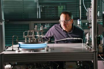
The new imaging technique takes advantage of a Cornell-patented fluorescence emission microscopy system and the natural fluorescence of certain bodily constituents.
Diagnoses of cancers and neurodegenerative diseases, such as Alzheimer’s disease, are two applications suggested by the researchers in their report in Proceedings of the National Academy of Sciences (PNAS , June 10, 2003), „Live tissue intrinsic emission microscopy using multiphoton-excited native fluorescence and second harmonic generation.“ The researchers predict that it should be possible to obtain endoscopic and laparoscopic images of tissues at the cellular level from deep within living animals, or even human patients, thus enabling a new form of optical biopsy.
The researchers have demonstrated the new imaging technique by making live-tissue intrinsic fluorescence scans of autopsy samples from the brains of patients with Alzheimer’s disease and by imaging mammary gland tumors in mice that serve as models of human cancer. Side-by-side comparison with conventional medical biopsy images of thin embalmed sections of the same organs reveals that the new method provides at least equal information, and in some cases contains additional diagnostic details not found in the conventional biopsies, which require invasive surgery.
Another advantage of live-tissue intrinsic emission imaging, the researchers say, is that the scans can be made through the surface of intact organs or body systems. By comparison, histopathology studies generally are performed on biopsy samples removed from subjects, then „fixed“ or embalmed and stained with labeling chemicals, which involves extended time delays.The Cornell-Harvard team incorporated a technology into the new imaging procedure called multiphoton microscopy, invented in 1989 by Watt W. Webb, Cornell’s S.B. Eckert Professor of Engineering and professor of applied physics, and Winfried Denk, now director of the Max-Planck-Institut für Medizinische Forschung Biomedizinische Optik, Germany.
Members of the imaging team included Webb, who also is director of the National Institutes of Health-funded Developmental Resource for Biophysical Imaging and Optoelectronics (DRBIO); Warren R. Zipfel, associate director of DRBIO, who designed and built the experimental system; Rebecca M. Williams, a DRBIO researcher who conducted many of the imaging tests; Richard Christie and Bradley T. Hyman of the Alzheimer Research Unit at Harvard Medical School’s Massachusetts General Hospital, who provided human brain tissue and diagnosed Alzheimer’s disease with the new imaging technique and the more traditional histopathology stain methods; and Alexander Yu Nikitin, assistant professor of pathology in the Department of Biomedical Science in Cornell’s College of Veterinary Medicine, who developed genetically engineered mouse models with humanlike cancers and then performed conventional diagnostic histopathology on the specimens.
„Multiphoton microscopy produces high-resolution, three-dimensional pictures of tissues with minimal damage to living cells,“ Webb explains. „Using a laser that produces a stream of extremely short, intense pulses, the probability that two or three interact with an individual biological molecule at the same time is greatly increased. When this occurs, their individual energies can combine. This cumulative effect is the equivalent of delivering one photon with twice the energy (or half the wavelength) in the case of two-photon excitation, or three times the energy (one-third the wavelength) in three-photon excitation,“ Webb notes.
The scanning laser microscope moves the focused beam of pulsed photons across a sample at a precise depth (plane of focus) so that cells above or below the plane are not affected, according to Webb. When repeated scans at different focal planes are „stacked,“ a three-dimensional picture emerges.
„Multiphoton microscopy is extremely well-suited to take advantage of the natural fluorescence [the ability to give off light under bombardment by radiant energy] of certain constituents in living tissue,“ says Zipfel. „Some amino acids [such as tryptophan and tyrosine] fluoresce naturally with ultraviolet light. Furthermore, many of the body’s vitamin derivatives, such as retinol and riboflavin, emit longer-wavelength fluorescence.“
Media Contact
Alle Nachrichten aus der Kategorie: Medizin Gesundheit
Dieser Fachbereich fasst die Vielzahl der medizinischen Fachrichtungen aus dem Bereich der Humanmedizin zusammen.
Unter anderem finden Sie hier Berichte aus den Teilbereichen: Anästhesiologie, Anatomie, Chirurgie, Humangenetik, Hygiene und Umweltmedizin, Innere Medizin, Neurologie, Pharmakologie, Physiologie, Urologie oder Zahnmedizin.
Neueste Beiträge

Merkmale des Untergrunds unter dem Thwaites-Gletscher enthüllt
Ein Forschungsteam hat felsige Berge und glattes Terrain unter dem Thwaites-Gletscher in der Westantarktis entdeckt – dem breiteste Gletscher der Erde, der halb so groß wie Deutschland und über 1000…

Wasserabweisende Fasern ohne PFAS
Endlich umweltfreundlich… Regenjacken, Badehosen oder Polsterstoffe: Textilien mit wasserabweisenden Eigenschaften benötigen eine chemische Imprägnierung. Fluor-haltige PFAS-Chemikalien sind zwar wirkungsvoll, schaden aber der Gesundheit und reichern sich in der Umwelt an….

Das massereichste stellare schwarze Loch unserer Galaxie entdeckt
Astronominnen und Astronomen haben das massereichste stellare schwarze Loch identifiziert, das bisher in der Milchstraßengalaxie entdeckt wurde. Entdeckt wurde das schwarze Loch in den Daten der Gaia-Mission der Europäischen Weltraumorganisation,…


